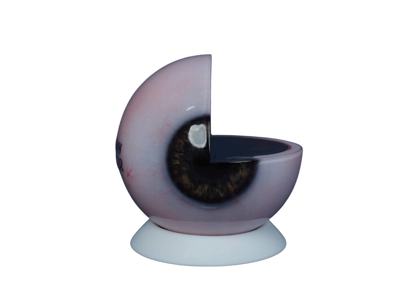
| Product Functions | Suitable for the display and teaching of the anatomical structure of the human eye, helping users master the complex and fine anatomical structures of the eyeball as well as their interrelationships and spatial positions. |
| Product Characteristics | 1. This model adopts a median sagittal and median horizontal 1/4 section, and the internal anatomical structures of the eye can be observed through its transparent material. 2. It highly accurately reproduces the anatomical structures of the eye, including the sclera, retina, cornea, pupil, iris, vitreous body, lens, zonule of Zinn, and retinal blood vessels. |

Specific Description

-
Address: 1001, 10th Floor, Building 1, No. 61, Huanhu South Road, Shilong Town,Dongguan,P.R.China
-
Service Hotline: +86 0769 82299001
-
Email: suhui@maxseas.com sales@maxseas.com
Copyright © 2025 Dongguan Maxseas Medical Technology Co., Ltd. All Rights Reserved.